IMAGING

See what fluorescence normally hides.

Intelligent Material converts near-infrared excitation into precisely engineered visible and near-infrared emission. Because the excitation wavelength lies above most endogenous fluorescence, the result can be exceptionally low background and high target-to-background imaging.

NIH lymphatic imaging figure showing white light, spectral imaging and single-shot imaging
980 NM EXCITATIONLOW BACKGROUND
THE ORIGIN STORY

National Institutes of Health research demonstrated in vivo lymphatic imaging.

A collaboration among the National Cancer Institute, NIH, University of Pennsylvania and Sunstone BioSciences demonstrated multi-color lymphatic imaging using rare-earth crystals excited at 980 nm.

Two engineered materials produced distinct emission signatures under the same excitation: a Yb/Tm formulation with emission at 470 and 800 nm, and a Yb/Er formulation with emission at 550 and 670 nm.

The study's central advantage was background. Under 980 nm excitation, the authors reported no detectable tissue autofluorescence in the spectral window of interest.

In vivo multiple color lymphatic imaging using upconverting nanocrystalsHisataka Kobayashi et al. Journal of Materials Chemistry, 19, 6481-6484. Authors included Christopher B. Murray, Josh Collins, G. Ajith Kumar and Howard Bell.DOI 10.1039/B910512C →
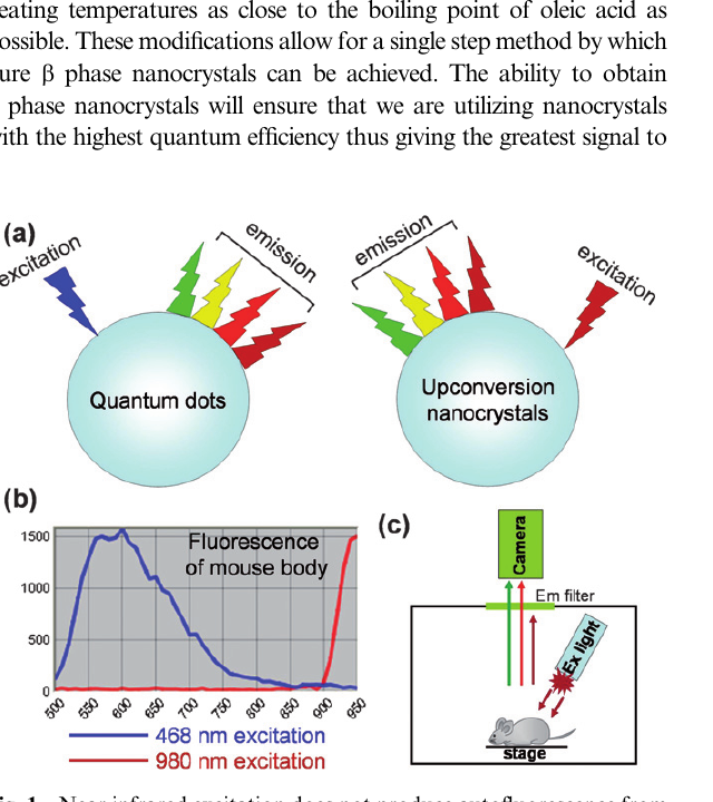
Published figure comparing excitation and background autofluorescence
Published NIH research: near-infrared excitation produced no detectable background from the mouse abdomen in the visible/NIR spectral window used in the study.
HOW IT WORKS

One excitation wavelength. Multiple engineered identities.

The crystal composition determines the optical response. A single 980 nm source can excite different Intelligent Material formulations, while the resulting emission wavelength becomes the information channel.

01 · Excite

Near-infrared energy in.

Illuminate the engineered rare-earth crystal at 980 nm, a wavelength that excites far less endogenous fluorescence than visible excitation in the reported experiment.

02 · Convert

The crystal changes the energy.

Rare-earth ions convert the incoming near-infrared excitation into shorter-wavelength optical emission.

03 · Program

Composition sets the response.

Yb/Tm and Yb/Er combinations created distinct optical signatures, demonstrating that the material can be engineered for different emission channels.

04 · Image

Read the emitted light.

The emitted signal can be detected spectrally or in a single shot, creating a direct optical readout from the physical material.

980 nm in
470 / 550 / 670 / 800 nm out
PUBLISHED EVIDENCE

Low background changed the imaging problem.

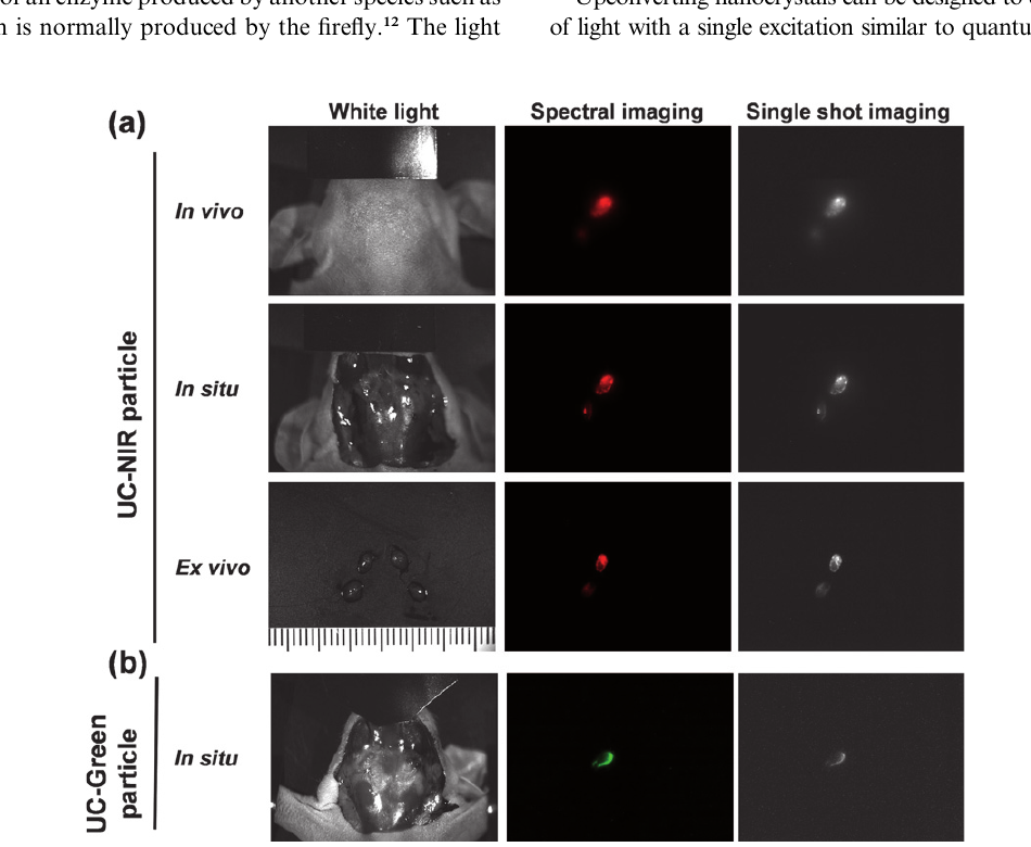
In the animal study, the NIR-emitting formulation depicted draining lymph nodes in vivo and in situ in all four mice. The authors reported that single-shot 800 nm images were comparable to spectrally unmixed images that required roughly 30 seconds of acquisition and post-processing.

4 / 4Animals showed draining lymph nodes with the NIR-emitting material in vivo and in situ.
980 nmSingle excitation wavelength used for both engineered materials.
~30 secAcquisition/post-processing time cited for the spectrally unmixed comparison images.
2 colorsSerial injections demonstrated multi-color lymphatic imaging using distinct emission channels.
Published in vivo, in situ and ex vivo lymph-node imaging figure
This was preclinical animal research, not a clinical efficacy study. The authors also noted that the approximately 20 nm crystal cores and roughly 50 nm coated particles were still larger than the size they considered optimal for in vivo imaging, and that smaller, brighter materials were under development.
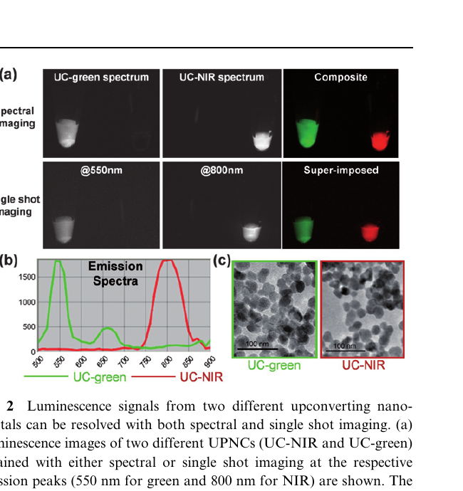
Published spectra and transmission electron microscopy figure
Two different material formulations were resolved by both spectral and single-shot imaging under the same 980 nm excitation.
MULTIPLEXING

Different crystals can carry different optical identities.

The NIH research is important beyond lymphatic mapping. It showed that two materials with different rare-earth compositions could be distinguished by their emission spectra while sharing the same excitation source.

That is the basis of a broader imaging platform: engineer a material for a specific target, give it a distinct optical response, then read multiple identities through one excitation architecture.

Same excitationDifferent emissionSingle-shot capable
TWO-COLOR LYMPHATIC IMAGING

Imaging can be spatial, spectral and temporal at once.

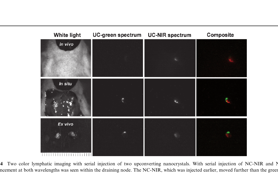
Serial injection of the two formulations produced enhancement at both wavelengths in draining lymph nodes. The earlier NIR-emitting material had moved farther through the lymphatic pathway than the later green-emitting material, demonstrating how location and material identity can be read together.

Because the optical response is engineered into the crystal, future systems can use wavelength, lifetime, intensity and other response dimensions as additional information channels.

Published two-color lymphatic imaging figure
Published two-color lymphatic imaging from the NIH research.
WHERE THIS CAN GO

Make the marker part of the medical workflow.

The published work established the optical principle. IMS is interested in extending that principle into practical imaging systems where the material itself carries a persistent, readable identity.

01

Lymphatic mapping

Low-background optical markers for locating draining lymph nodes and following lymphatic pathways.

02

Image-guided surgery

Materials designed to help localize tissue, biopsy sites or surgical targets before, during or after a procedure.

03

Multiplex molecular imaging

Distinct rare-earth compositions can provide multiple optical channels under a shared excitation source.

04

Persistent markers

Engineered materials can support concepts where the physical marker remains detectable over time rather than relying only on transient fluorescent dyes.

IMAGING COLLABORATION

What should Intelligent Material help you see?

IMS develops the material and intellectual property. We work with medical, research and instrumentation partners to adapt the optical response to a specific imaging problem.

Work with IMS →